Knee Tendon Diagram : Knee Injuries And Degeneration Anova Irm Germany / Its complexity and its efficiency is the best example of god's creation.
byAdmin-
0
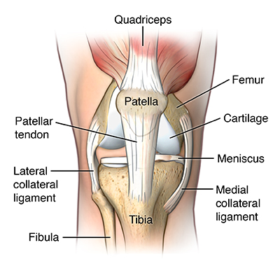
Knee Tendon Diagram : Knee Injuries And Degeneration Anova Irm Germany / Its complexity and its efficiency is the best example of god's creation.. Knee tendons diagram (page 1). The knee is designed to fulfill a number of functions: It is held in place by a ligament at the bottom and a tendon on top. The knee ligaments connect the thigh and shin bones (femur & tibia) and work together to control how the knee moves to keep it stable and prevent injury. The anatomy of the knee consists of bones, muscles, nerves, cartilages, tendons and ligaments.
Diagram of a catheter in the neck. Bertram zarins of the mass general hospital sports medicine service has prepared this animation to educate patients about the anatomy of the ligaments wh. Furthermore, there are several individualized. The severity of these symptoms depends on which ligament has been torn. The two peroneal tendons in the foot run side by side behind the outer ankle bone.
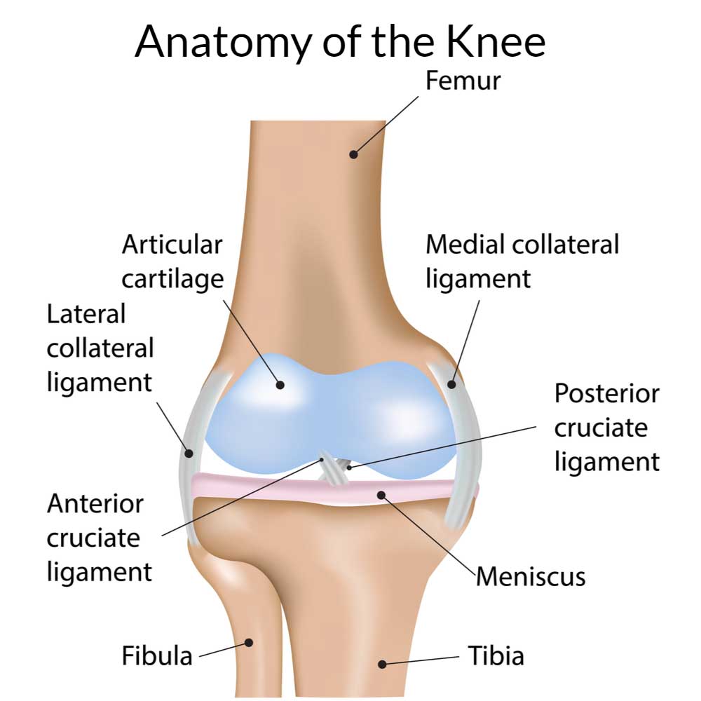
Patellar Tendonitis Jumper S Knee Johns Hopkins Medicine from www.hopkinsmedicine.org Damage in even one part can hinder the functioning of the knee. The knee joint is most significantly affected by two major muscle groups: Furthermore, there are several individualized. The knee ligaments connect the thigh and shin bones (femur & tibia) and work together to control how the knee moves to keep it stable and prevent injury. The patella tendons surround the kneecap and the quadriceps tendons are toward the back of the knee and leg. Acl & pcl found in the middle of the joint. The knee consists of three bones: In the knee, they give stability and strength to the knee joint as the bones and cartilage of the knee have very little stability on their own.
It is disabling pain and it gets worse with extending and standing and walkig.
The anterior cruciate ligament prevents the femur from sliding backward on the tibia (or the tibia sliding forward on the femur). The largest joint in the body, the knee moves like a hinge, allowing you to sit, squat, walk or jump. Knee diagram tendons was posted in may 29, 2015 at 4:57 pm. Bertram zarins of the mass general hospital sports medicine service has prepared this animation to educate patients about the anatomy of the ligaments wh. We have a collection of human body muscle diagram to help you learn more about the topic. Then next one, further down, looks at pain behind the knee. Ligaments are elastic bands of tissue that connect bones to each other and provide stability and strength to the joint. Ankle tendon anatomy, hamstring tendon, knee ligament anatomy, knee tendon pain, knee tendonitis, lateral collateral ligament, patella tendon anatomy, patellar tendon, foot, ankle tendon anatomy, hamstring tendon, knee ligament anatomy, knee tendon pain, knee tendonitis, lateral. A tendon connects the muscle to the bone. Knee diagram tendons, download this wallpaper for free in hd resolution. They are attached to the femur (thighbone), tibia (shinbone), and fibula (calf bone) by fibrous tissues called ligaments. Muscles propel the knee joint back and forth. Knee tendon diagram / knee anatomy the basics the knee expert / around the knee there are two types of tendons.
Knee pain could be the result of a problem with any one of these components, or a combination of several. The largest joint in the body, the knee moves like a hinge, allowing you to sit, squat, walk or jump. Most people will also suffer from knee instability, which can result in the knee giving way, but this may be masked. Tendons are the connection between bones and muscles tendon diagram. The knee ligaments connect the thigh and shin bones (femur & tibia) and work together to control how the knee moves to keep it stable and prevent injury.
Knee Anatomy Orthopedic Knee Specialist Manhattan New York City Ny from rileywilliamsmd.com It connects the thigh bone to the shin bone. Ankle tendon anatomy, hamstring tendon, knee ligament anatomy, knee tendon pain, knee tendonitis, lateral collateral ligament, patella tendon anatomy, patellar tendon, foot, ankle tendon anatomy, hamstring tendon, knee ligament anatomy, knee tendon pain, knee tendonitis, lateral. The knee consists of three bones: Anterior cruciate ligament (acl) is the most commonly injured knee ligament. Mcl & lcl found either side of the knee. Knee tendon diagram / knee anatomy the basics the knee expert / around the knee there are two types of tendons. Ab 50€ portofrei, versand innerhalb 24h, 100 tage retoure, über 1 mio. Diagram of the ankle bones.
Acl & pcl found in the middle of the joint.
Then next one, further down, looks at pain behind the knee. Tendons are the connection between bones and muscles tendon diagram. We have a collection of human body muscle diagram to help you learn more about the topic. Anterior cruciate ligament (acl) is the most commonly injured knee ligament. When the muscle contracts, the tendons are pulled, and the bone is moved. Ligaments join the knee bones and provide stability to the knee: Each of the 6 sections ( bones, connective tissue 1, connective tissue 2, deep muscles, muscles & skin) can be opened up, rotated left or right and viewed more closely. In the knee, they give stability and strength to the knee joint as the bones and cartilage of the knee have very little stability on their own. The knee consists of three bones: The muscles that affect the knee's movement run along the thigh and calf. Diagram of knee joint showing mcl. Diagram of inside the body. Knee tendon diagram / knee anatomy the basics the knee expert / around the knee there are two types of tendons.
Furthermore, there are several individualized. The knee ligaments connect the thigh and shin bones (femur & tibia) and work together to control how the knee moves to keep it stable and prevent injury. Ligaments are elastic bands of tissue that connect bones to each other and provide stability and strength to the joint. Jumper's knee is diagnosed by taking a medical history and doing a physical exam. The knee is the joint where the bones of the lower and upper legs meet.
Patellar Tendon Anatomy Origin Insertion Function Kenhub from thumbor.kenhub.com Knee pain could be the result of a problem with any one of these components, or a combination of several. Diagram of knee joint showing mcl. The four main ligaments in the knee connect the femur (thighbone) to the tibia (shin bone), and include the following: Tendons are the connection between bones and muscles tendon diagram. The muscles that affect the knee's movement run along the thigh and calf. The knee ligaments connect the thigh and shin bones (femur & tibia) and work together to control how the knee moves to keep it stable and prevent injury. This tendon connects the patella (kneecap) to the tibia. A dislocated kneecap is yet another common knee condition.
A tendon is a band of tissue that connects a muscle to a bone.
The two peroneal tendons in the foot run side by side behind the outer ankle bone. Then next one, further down, looks at pain behind the knee. This first knee pain diagnosis chart focuses on pain at the front of the knee. This tendon connects the patella (kneecap) to the tibia. Bertram zarins of the mass general hospital sports medicine service has prepared this animation to educate patients about the anatomy of the ligaments wh. Hand tendons diagram, picture of hand tendons diagram. The knee ligaments connect the thigh and shin bones (femur & tibia) and work together to control how the knee moves to keep it stable and prevent injury. A diagram of the knee, including ligaments. Hochwertige kletterseile für dein outdoor abenteuer! The knee is the joint where the bones of the lower and upper legs meet. Diagram of inside the body. Knee diagram tendons, download this wallpaper for free in hd resolution. Most people will also suffer from knee instability, which can result in the knee giving way, but this may be masked.
It is disabling pain and it gets worse with extending and standing and walkig tendon diagram. Diagram of the ankle bones.